“Leaky Valve Disease” of Older Dogs

Degenerative Atrio-Ventricular (Mitral and Tricuspid Regurgitation) Valve Disease

What dogs get this disease? 

This very common disease affects dogs usually after the age of 8 years. Small breed dogs are most commonly affected. Some of the breeds include Cavalier King Charles Spaniels, Miniature poodles, Shih Tzu, Maltese, Chihuahua, Cocker Spaniels, Miniature Schnauzers, Dachshunds, Whippets, and Pomeranians. This disease can also be seen in some larger breeds and mixed breed dogs.

CAUSE: 

There are 4 valves in the heart and the most commonly affected with the greatest leakage is the mitral valve. This valve separates the left ventricle from the left atrium. Normally this valve closes when the heart contracts and this closure prevents blood from going back into the atrium so that normal all the blood goes to the body. When the valve leaks the blood goes backwards into the left atrium. The flow of blood going the wrong way is called “regurgitation”. Therefore, this disease often is called mitral regurgitation. (If the valve between the right ventricle and right atrium also is affected the term tricuspid regurgitation is used.) Eventually the left atrium enlarges followed by fluid accumulation in the lungs because of too much volume and pressure. The valve leaks because it has “degenerated”. The normal structural integrity of the leaflets of the valve is lost. The valve is composed of elastic and collagen tissue with another substance called the spongiosa. In the older dog the tissues (elastic and collagen fibers) that give support and resiliency to the valve weaken and the spongiosa gets thicker. The thickened valve “flops” more (think about the loss of similar tissues in skin and the difference between a young and old person!). As a result of these changes the thickened valve does not close properly. So it leaks.

CLINICAL SIGNS: 

Many dogs in the early stages have no clinical signs or they are so subtle that owners just believe that a dog is “slowing down” because of age or other common aging changes such as arthritis. Clinical signs do include exercise intolerance, coughing, trouble breathing, increased breathing rate, collapse, or weakness.

DIAGNOSIS: 

Virtually all dogs with clinically important mitral and tricuspid regurgitation will have a cardiac murmur heard when the chest is listened to with a stethoscope. This is auscultation of the chest. Veterinarians can hear a murmur long (months to years) before clinical signs are noticed. In contrast, most frequently, early degeneration of the valve does not cause a murmur because the amount of leaking is not enough to cause a sound that can be heard on the surface of the body as it originates within the heart. Radiography Radiographs which are made with x-rays provide information regarding the presence of fluid in the lungs and the size and shape of the silhouette of the heart. As disease progresses the heart size is seen to increase as shown in radiographs. Echocardiography An important diagnostic tool to fully characterize the structure and function of the valves and heart is ultrasound of the heart known as echocardiography. This test permits the examination not only of the muscle and valves, but also of the blood flow (Doppler echocardiography). Therefore, the changes to the heart are measured and the amount of blood “going the wrong way” can be semi-quantified. NT-proBNP. This is a blood test that may indicate advanced heart failure when a large amount of regurgitation is present. Electrocardiography An electrocardiogram (ECG) may be preformed to further characterize dogs with AV valve degeneration; however, this test is usually not as important as the physical examination, radiograph, and echocardiogram. Other tests may be ordered to determine the status of other body systems and to insure that all are functioning adequately for the clearance of the drugs that will be used to treat the disease process.

TREATMENT: 

Numerous drugs are used to treat this disease. It is vital that the drugs selected and doses are titrated to the need of each dog. Early disease is not treated the same as advanced disease. This is no different than the variation in treating cancers at different stages. Because valve degeneration is slowly progressive, treatment can change over the course of months or even years. Drugs that are commonly used are diuretics (e.g. furosemide), vasodilators (e.g. enalapril, benazapril, pimobendan), positive inotropes (e.g. pimobendan, digoxin) and other drugs to address specific problems like high pressure in the vessels of the lungs. Unfortunately, treatment of this disease is not curative, but palliative. Further research is really required to give knowledge for therapeutic intervention early that will stop the progression of the degeneration of the valve.

PROGNOSIS:

 The prognosis depends on the severity of the disease which is determined by when it is identified. Many dogs identified early live for many years, while others with clinical signs may only live a few months. The diagnostic tests will not only give the diagnosis, but the staging of the disease for each dog.

How should an affected dog be monitored? 

The frequency and intensity of the monitoring depends on each patient, the stage of the disease, the response to treatment, and the status of other body systems.

Read More…

Hypertrophic Cardiomyopathy (HCM)

CAUSE: 

PureVeter | After Hours Urgent Care Clinic for Pets | Jacksonville FL

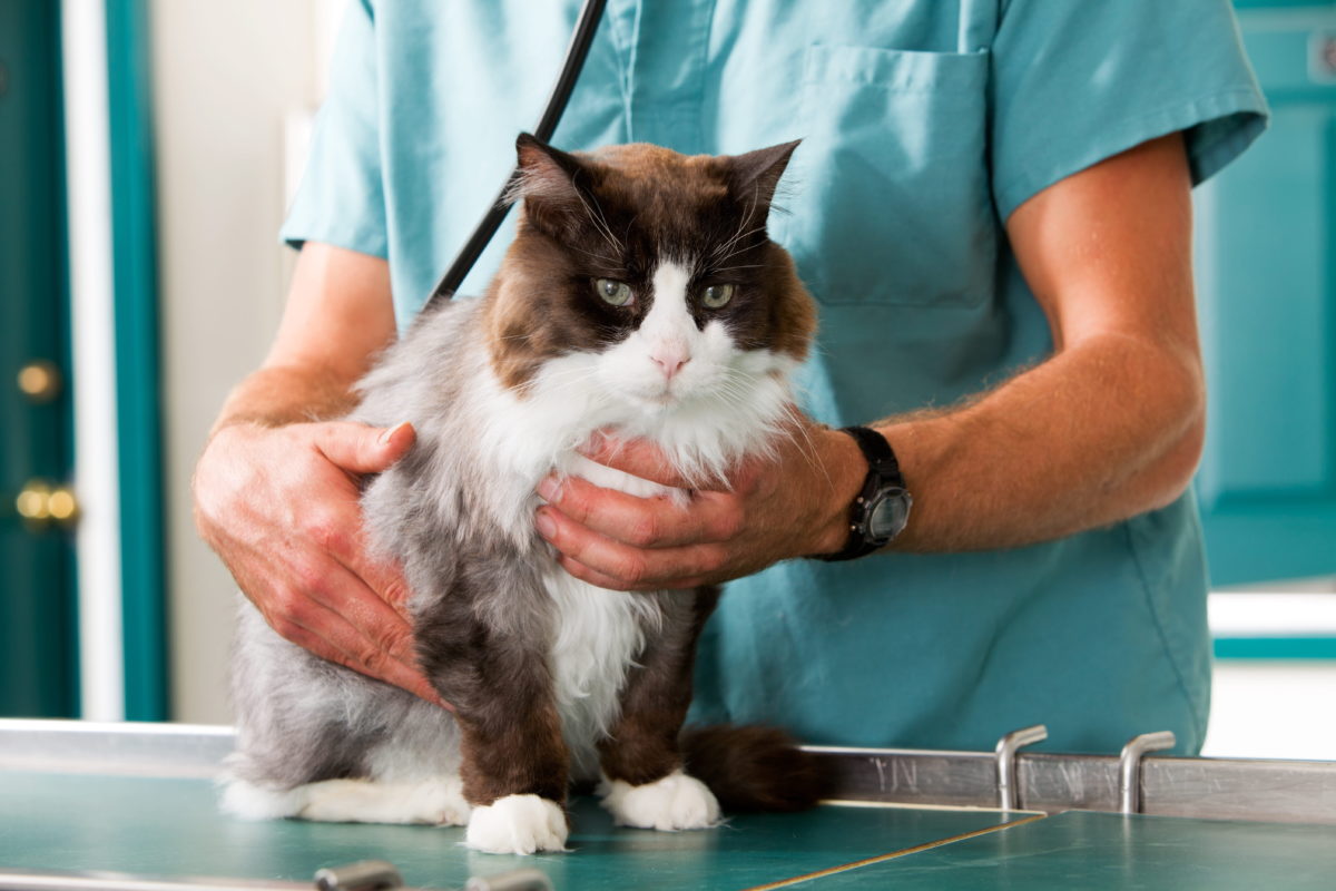
Hypertrophic cardiomyopathy (HCM) is the most commonly diagnosed cardiac disease in cats. Characterized by regional or diffuse thickening of the walls of the ventricle (the primary “pump” muscle of the heart), HCM has been diagnosed in cats as young as 4 months old and as old as 16 years old. Although the definitive cause of feline HCM has not been identified, its prevalence within certain breeds (i.e. Maine Coon cats, Ragdolls) has prompted speculation that at least some forms of HCM are genetic in origin. The finding of mutations in an important cardiac protein called myosin binding protein C in affected lines of Maine Coon and Ragdoll cats supports a heritable, genetic component of HCM in these breeds.

Thickening of the walls of the ventricle is associated with a decreased ventricular chamber volume and abnormal ventricular relaxation (diastolic function) in cats with HCM. Since the amount of blood pumped by the heart per minute (cardiac output) is the product of the amount of blood ejected per contraction (stroke volume) and the heart rate in beats per minute, this decreased chamber volume (and subsequent stroke volume) results in an increased heart rate (tachycardia) as a reflex mechanism to maintain cardiac output and blood pressure. Although this reflex increase in heart rate may maintain normal blood pressure in the short term, it is associated with an increased consumption of oxygen by the heart muscle, to the extent that oxygen demand may exceed supply. This scenario may result in an energy starved heart muscle, with subsequent heart cell death and worsening function. Another consequence of an increased heart rate is that the ventricle has less time to fill between contractions, further diminishing stroke volume and promoting a vicious cycle of reflex tachycardia, decreased time for ventricular filling, and so on. This decreased left ventricular filling also promotes stasis of blood in the left atrium (chamber just before the left ventricle), which ultimately contributes to the development of clinical signs (see below).

CLINICAL SIGNS: 

Many cats with HCM present without overt signs of illness. In other cases, signs of congestive heart failure including labored or rapid breathing, open-mouth breathing, and lethargy are evident. These signs occur when fluid accumulates in lung tissue (pulmonary edema) or around the lungs (pleural effusion) secondary to elevation of left atrial pressure. A potentially devastating cclud of HCM is thromboembolism. Thromboembolism refers to the development of a clot in the heart (promoted by left atrial enlargement), with ejection of the clot to the systemic circulation. When the clot lodges in the peripheral circulation, it may obstruct blood flow to the region of the heart supplied by the blocked vessel. The site of thromboembolism most commonly observed in cats with HCM is the distal aorta (termed a saddle thrombus), and clinical signs of hind limb paralysis and acute pain in the hind limbs may be observed. Thromboembolism is a poor prognostic indicator in cats with HCM.

DIAGNOSIS:

 HCM is diagnosed by echocardiography, which shows the characteristic thickening of the left ventricular walls and decreased chamber volume of the left ventricle. Evaluation of the left atrium for dilation and/or the presence of a thrombus are also achieved using this modality. Since hyperthyroidism and hypertension may also cause left ventricular thickening, these diseases must be ruled out prior to arriving at a diagnosis of HCM. Thoracic radiography may be useful to evaluate pulmonary (lung) status and to rule out pleural effusion. Electrocardiography may be useful to characterize heart rate and to rule out cardiac arrhythmias.

TREATMENT: 

Treatment goals for feline HCM include controlling heart rate, alleviating pulmonary congestion, removing pleural fluid (if present), and decreasing the likelihood of thromboembolism. These goals are addressed by the administration of medications that may be delivered by injection in emergent situations or orally in more stable patients. Some drugs (i.e. nitroglycerine) may be applied to the skin for absorption.

PROGNOSIS: 

The prognosis for cats with HCM is quite variable. Cats without clinical signs may survive for years, although the disease is most commonly progressive. Poor prognostic indicators include the presence of congestive heart failure, thromboembolism, and hypothermia (low body temperature). In spite of the fact that HCM may decrease the life span of affected cats (sometimes significantly so), medical therapy can improve the quality of life and longevity of cats affected with this common disease.

Read More…